Get the latest news, application notes, and upcoming webinars from XGen Bio Inc.
- About
- About XGen
- Our team
- Follow Us
- Youtube
![]()
CONTACT US
Copyright© 2025, Xgen. All rights Reserved.
Terms & Conditions | Privacy Policy | Cookie Policy
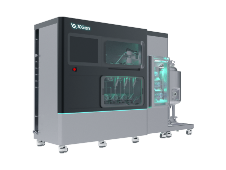
INano™ L+
Rapid Nanomedicine Preparation System
INano™ Optimux
Medium-Scale Formulation (Reusable System & GMP Compliant)
INano™ S
Commercial LNP Manufacturing System

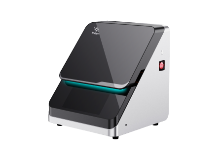
XNano™ HT-Smart
End-to-End High-Throughput mRNA/LNP Screening Workstation
XNano™ PCV
LNP Manufacturing System For Emerging Applications
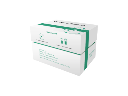
mRNA-LNP kit
Cell Transfection Kit
Application Kit
Organ-specific Targeting Kit
Validation Kit

DNA/Protein-LNP Kit
DNA-LNP Kit
Protein-LNP Kit
|
This study explores a novel strategy that utilizes apolipoprotein fusion to achieve spontaneous antibody functionalization of mRNA lipid nanoparticles (LNPs). This research demonstrates that this method significantly enhances the targeted delivery efficiency and bioavailability of LNPs, while maintaining their stability and biocompatibility. This innovative LNP functionalization strategy holds great potential for cancer therapy and other medical applications that require precise delivery.
Materials and Methods LNP Preparation: Standard LNP preparation techniques were used, including the mixing of ionizable lipids, helper lipids, cholesterol, and PEGylated lipids. GrAb were then incorporated onto the LNP surface via incubation. Apolipoprotein Fusion: Using genetic engineering techniques, apolipoproteins were fused with targeted antibodies for expression. Characterization and Testing: The prepared LNPs were characterized using a variety of physical and chemical methods, such as particle size analysis, Zeta potential measurement, gel electrophoresis, and cytotoxicity assays. Flow cytometry and fluorescence microscopy were employed to observe the intracellular delivery of LNPs. Animal Experiments: In mouse models, the in vivo delivery efficiency and biological safety of the LNPs were assessed. The targeted delivery and bioavailability of the LNPs were evaluated by monitoring antibody levels in serum and tissue distribution.
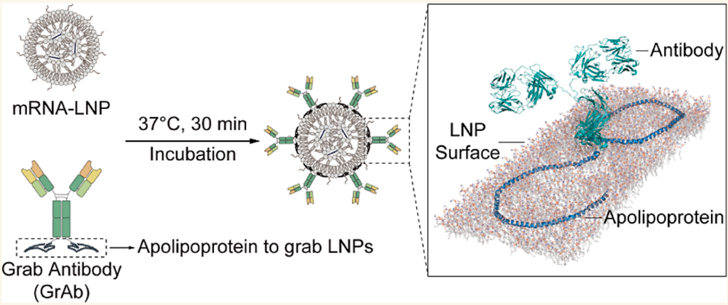
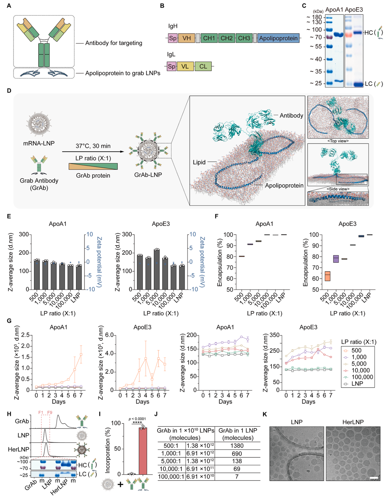
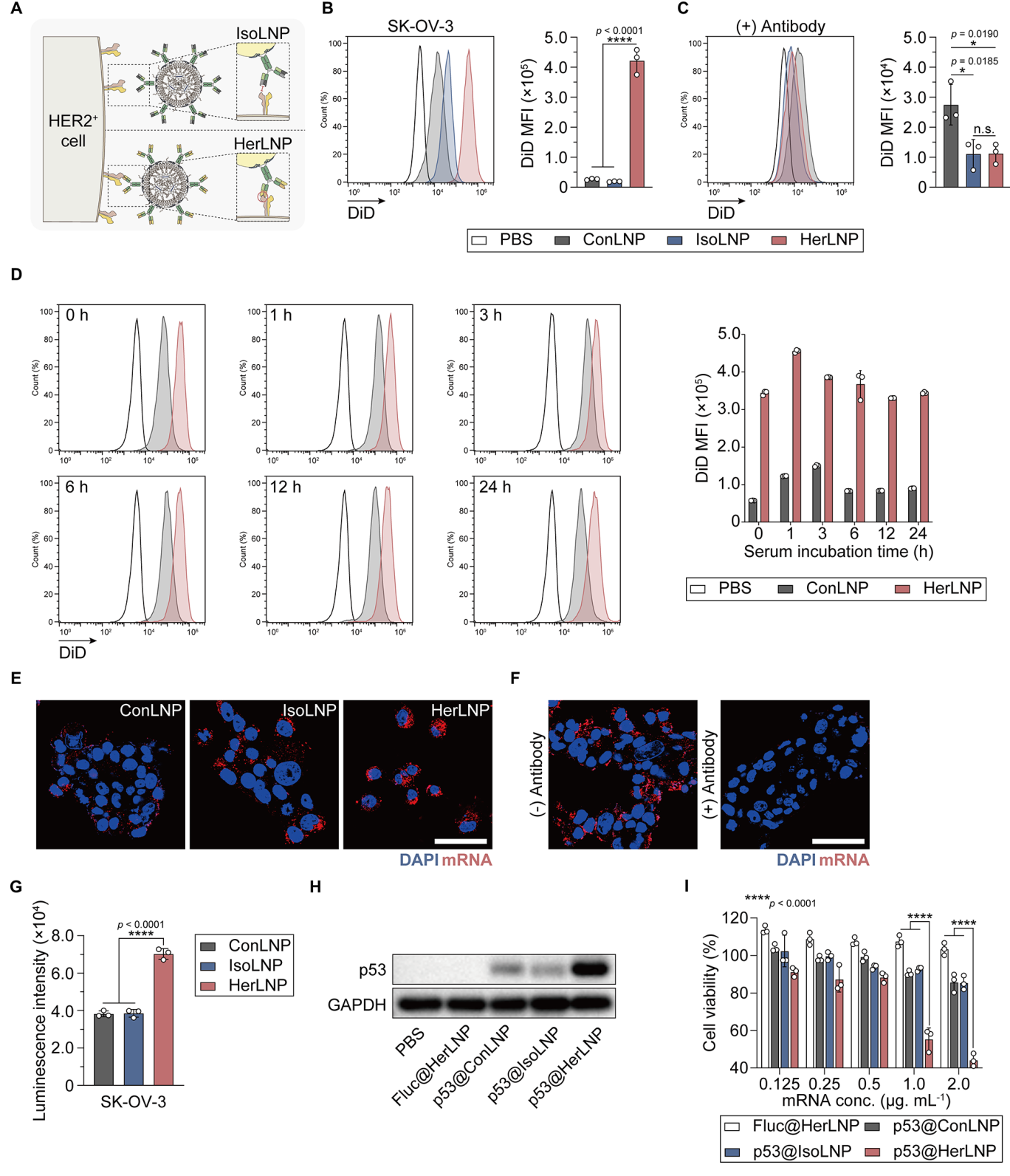
Results Preparation and Characterization of Antibody-Functionalized LNPs The researchers successfully constructed fusion plasmids encoding the antibody N-terminal signal sequence, heavy chain, and apolipoprotein, as well as a plasmid encoding the antibody light chain with an N-terminal signal sequence. By co-expressing these plasmids, we obtained the engineered antibody (GrAb) with the apolipoprotein fusion. The GrAb was then incubated with LNPs at 37°C for 30 minutes, allowing the strong lipid-binding affinity of apolipoproteins domain to spontaneously anchor the GrAb on the surface of the LNPs. Dynamic light scattering (DLS) results showed that the Z-average particle size and Zeta potential of GrAb-LNPs did not significantly differ from those of bare LNPs, indicating that the antibody functionalization process did not notably alter the particle size or surface charge. Additionally, mRNA encapsulation efficiency was assessed using the RiboGreen fluorescence assay. The results showed that the mRNA encapsulation efficiency of GrAb-LNPs was comparable to that of bare LNPs, confirming that the antibody functionalization process did not affect the mRNA encapsulation capacity of the LNPs. Cell-based studies demonstrated that GrAb-LNPs significantly enhanced uptake by target cells, indicating the successful targeting and functionalization of LNPs with the antibody. These findings validate the effectiveness of the apolipoprotein fusion method in functionalizing LNPs without compromising their structural integrity or mRNA encapsulation efficiency.
Figure 1: Construction and Characterization of GrAb-LNP Platform
Figure 2: In Vitro Targeting Capability of GrAb-LNP Platform
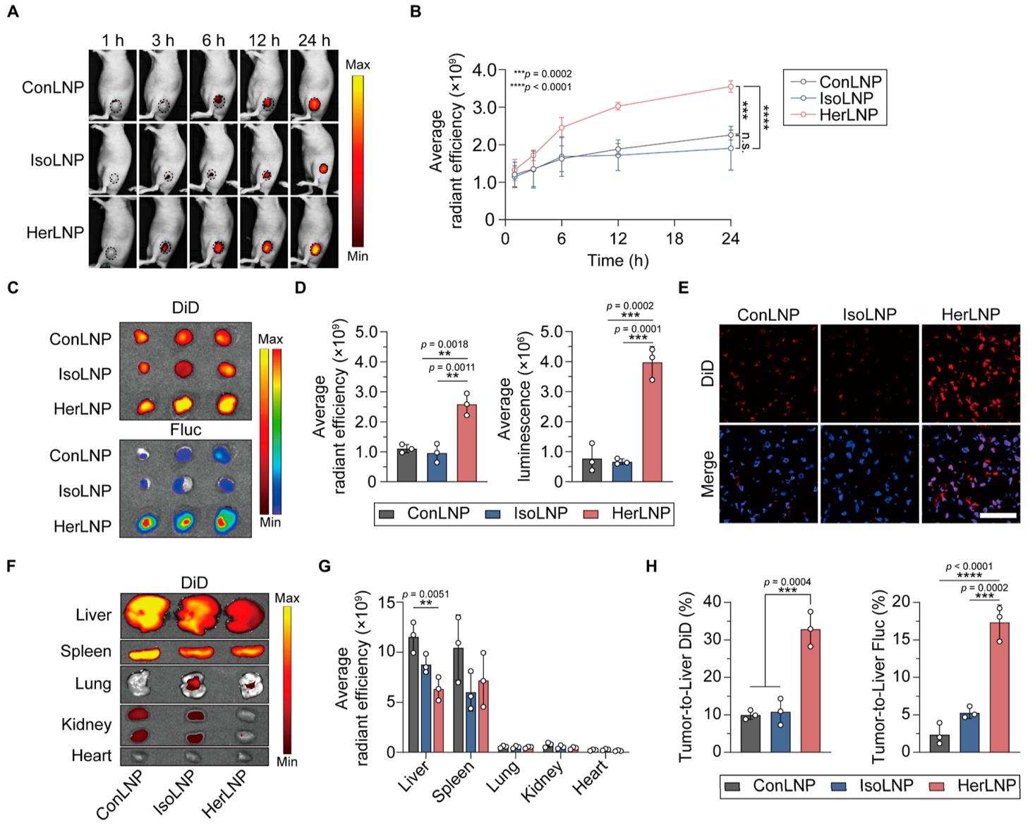
Targeted Delivery Efficiency of GrAb-LNPs To evaluate the targeted delivery efficiency of GrAb-LNPs, the researchers utilized a HER2-positive SK-OV-3 tumor mouse model. DiD-labeled LNPs containing luciferase (Fluc) mRNA were intravenously injected into the mice, and near-infrared fluorescence (NIRF) imaging was performed at various time points. The results showed that, compared to bare LNPs, HerLNPs (LNPs functionalized with GrAb targeting HER2) exhibited significantly enhanced fluorescence signals at the tumor site, with the signal gradually increasing over time. This indicates that HerLNPs efficiently deliver mRNA to the tumor site. These findings suggest that GrAb-LNPs, especially when functionalized with HER2-targeting antibodies, have significant potential for targeted mRNA delivery in tumor therapy.
Figure 3: In Vivo Targeting Capability of GrAb-LNP Platform
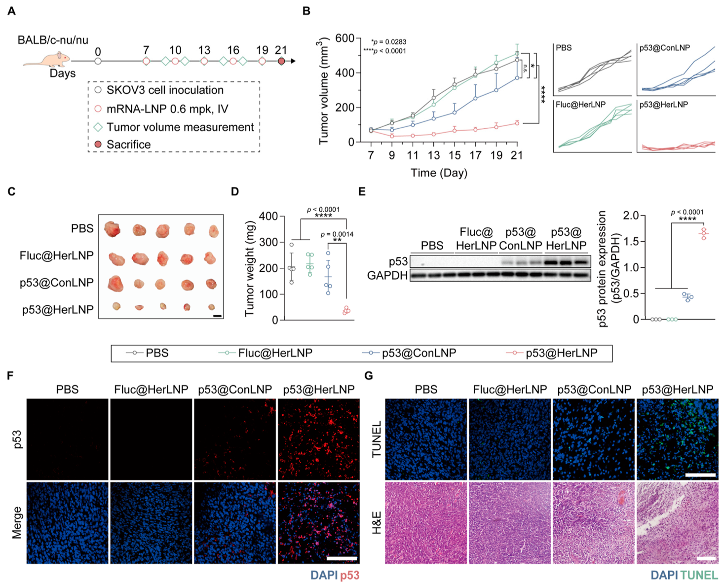
Antitumor Effect of GrAb-LNPs To evaluate the antitumor effect of GrAb-LNPs, the researchers intravenously injected HerLNPs containing p53 tumor suppressor gene mRNA into HER2-positive tumor-bearing mice. The results showed that, compared to the bare LNPs or the control group, the HerLNP-treated mice exhibited significantly reduced tumor volumes, with some tumors completely regressing. This demonstrates that HerLNPs can efficiently deliver p53 mRNA into tumor cells, upregulating tumor suppressor expression and thereby inhibiting tumor growth. These findings highlight the potential of GrAb-LNPs as a highly effective targeted therapy for cancer, particularly in HER2-positive tumors.
Figure 4: In Vivo Antitumor Effect Evaluation of HER-2 Targeted p53 mRNA/LNPs
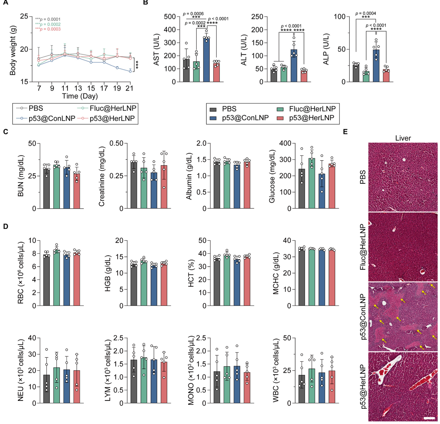
Biocompatibility and Safety of GrAb-LNPs To assess the biocompatibility and safety of GrAb-LNPs, they performed histological analysis. The results showed that, compared to mice treated with bare LNPs (lacking antibody modification), mice treated with HerLNPs showed no structural damage or toxicity in major organs, including the lungs, spleen, kidneys, and heart. This indicates that GrAb-LNPs exhibit good biocompatibility and safety. These findings provide confidence in the potential clinical use of GrAb-LNPs for safe and effective therapeutic mRNA delivery.
Figure 5: In Vivo Safety Evaluation of GrAb-LNPs
Discussion In this study, the researchers successfully developed and characterized self-targeted antibody-functionalized LNPs through apolipoprotein fusion. The results demonstrated a significant improvement in delivery efficiency and biosafety. The fusion of apolipoprotein not only enhanced the targeting ability of LNPs but also increased their stability and biocompatibility through its natural lipid-binding properties. This strategy provides a new solution for mRNA vaccine and therapeutic delivery systems with broad applications. However, further studies are needed to elucidate the specific mechanisms underlying the apolipoprotein fusion process to optimize LNP preparation and delivery. Additionally, while this study achieved positive results in mouse models, further validation in other animal species and human trials is necessary to fully confirm its safety and efficacy.
References: Park W, Choi J, Hwang J, et al. Apolipoprotein Fusion Enables Spontaneous Functionalization of mRNA Lipid Nanoparticles with Antibody for Targeted Cancer Therapy[J]. ACS nano, 2025. |
Get the latest news, application notes, and upcoming webinars from XGen Bio Inc.
![]()
CONTACT US
Copyright© 2025, Xgen. All rights Reserved.
Terms & Conditions | Privacy Policy | Cookie Policy